Lameness in horses comes in two basic forms: primary and secondary.
Primary asymmetry occurs as a consequence of an event that originates independent of other pre-existing lameness. Trauma, breed, age, and poor conformation could all play a role in the development of primary lameness. Identifying the primary gait deficit(s) is the chief goal of the observer, because this is where treatment will eventually be directed. Secondary lameness, on the other hand, manifests as a consequence of one or more pre-existing gait deficits elsewhere in the horse. It can be genuine (as in cases of associated and compensatory issues) or artificial (as in the case of referred asymmetry). The relationship between primary and secondary lameness is unidirectional, meaning secondary lameness would not exist without the presence of a primary underlying problem. This is an important concept when considering the fact that permanent resolution of secondary lameness would, at least in part, demand resolution of its primary counterpart. So, as long as a primary problem exists, the potential for secondary lameness is not far behind.
On the other hand, secondary lameness may or may not coexist with primary lameness. A single primary lameness with no secondary elements would be classified as simple. Simple lameness is the most basic form, since examination, diagnosis, and treatment are all directed toward a single anatomic region of the horse.
In cases of complicated lameness, more than anatomic region is involved. Differentiating regions that require special attention from those that don’t is one of the primary objectives of the adept observer. All primary issues will require accurate diagnosis and treatment in order to reestablish the horse’s performance. Depending on the duration and nature of secondary lameness, however, exclusive treatment may or may not be necessary. In many instances, secondary issues will spontaneously resolve once the primary issue has been successfully addressed. The smarter approach, therefore, is to identify and treat primary problems first.
Consider the analogy of your car’s front-end alignment and how it affects tire wear. Poor alignment would be considered a primary issue. Accelerated tire wear would be expected to occur secondary to poor alignment. The expensive and time-consuming application of new tires might improve your car’s performance in the short term, but the persistence of poor alignment will repeatedly result in premature tire depletion. The appropriate course of action is unidirectional: fix the car’s alignment first, then evaluate the status of the tires to determine if and when replacement is necessary. Successful management of a horse’s soundness over the long term requires a similar approach.
Regular Observation Means Fewer Complicated Cases
Those of us who regularly and carefully observe our horses in motion will see fewer complicated cases because we are more likely to detect abnormalities soon after their onset and before additional primary or secondary issues have time to develop. Unless it’s due to a common traumatic event, it is relatively rare for two separate, unrelated problems to occur simultaneously. In this way, regular observation actually simplifies the process of evaluation by reducing the likelihood of complicated and secondary lameness.
Even so, the majority of lameness cases are complicated. In order to diagnose and treat appropriately, we must first categorize each component of the horse’s lameness as primary or secondary. Although this task may seem daunting to the casual observer, there are some basic guidelines that will help. Consider the following:
- All lame horses have at least one primary component that is contributing to their altered movement. That said, most lame horses have only one primary component. Multifactorial lameness is a relatively uncommon form of complicated lameness.
- There is little correlation between the degree of lameness and its primary/secondary designation. A severe lameness is not always a “primary” one, since secondary lameness is often more visibly obvious than its underlying primary one.
- Forelimb lameness is more likely to be secondary to hind limb lameness than vice versa. Since the forelimbs normally experience the majority of the horse’s weight, very slight hind limb lameness may be enough to significantly influence forelimb load and action. By contrast, marked forelimb lameness is typically required to generate corresponding repercussions behind.
- When forelimb and hind limb asymmetries coexist:
— If the hind limb component is worse, it is more likely to be primary.
— If the forelimb component is considerably worse (by more than one grade), it is more likely to be primary.
— If the severity of the two is comparable (within one grade of each other), the hind limb component is more likely to be primary. - Back and neck issues are more likely to be secondary to limb lameness than vice versa. In fact, secondary sacroiliac (SI) and back pain are frequently associated with chronic and/or moderate hind limb lameness in horses.
Common Forms of Secondary Lameness
Compensatory lameness occurs as a result of excessive stress experienced by tissues in one part of the body in response to a primary issue elsewhere in the body. Accordingly, compensatory problems don’t typically occupy the same region of the body as their primary source. In most cases, they occur in a limb experiencing abnormal weight-bearing forces and/or an abnormal flight path as a result of a primary issue in a separate limb. Compensatory issues can develop from chronic overloading as the horse attempts to protect the primarily affected limb. Compensatory lameness might be observed in the following cases, as examples:
- A horse that developed laminitis in one forelimb as a result of chronic severe lameness in the other (contralateral) forelimb.
- A horse that developed proximal suspensory desmitis in a forelimb due to chronic overloading in an effort to protect the contralateral hind limb (of the same diagonal pair).
The following characteristics are typically representative of compensatory lameness:
- Compensatory lameness in the forelimb generally occurs secondary to primary lameness in the contralateral forelimb or hind limb. For example, primary issues in the right hind (RH) and/or right front (RF) limbs could precipitate compensatory lameness in the left forelimb (LF).
- Compensatory lameness in the hind limb usually occurs secondary to a primary weight-bearing lameness in the contralateral (other) hind limb. For instance, we would expect secondary symptoms associated with severe weight-bearing lameness of the RH limb to manifest most readily in the LH limb. Unless chronic and severe, it would be unlikely for a forelimb lameness to precipitate compensatory lameness in a hind limb.
Associated Lameness
Associated lameness, though secondary, does not generate complicated asymmetry and, therefore, does not need to be designated until the veterinary diagnostic phase of examination.
Causes of associated gait deficits generally reside in the same region as the primary source of lameness. Their presence further alters the horse’s gait and may affect both the degree and nature of abnormal movement. Variations in the amount of local inflammation and/or modifications in motion or weight bearing can precipitate associated problems within the affected limb.

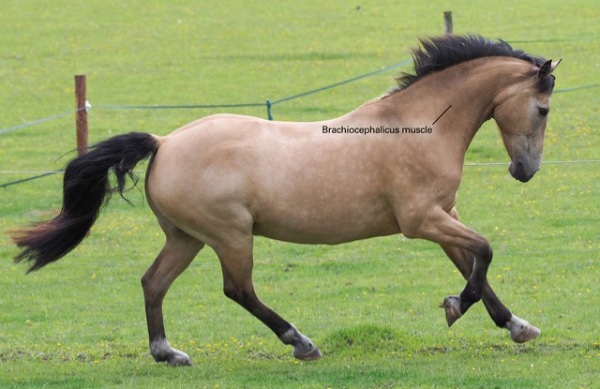
The brachiocephalicus muscle.
Examples of associated lameness might include the following:
- Greater trochanteric bursitis (also known as “whorlbone”) is commonly associated with lower hock pain. Inflammation within the greater trochanteric bursa often occurs as a result of chronic excessive pelvic limb adduction (i.e. pulling the limb underneath the center of the body) during movement. This motion results in increased strain of the middle gluteal muscle and its associated tendon. Excessive limb adduction is, in turn, a gait characteristic classically associated with distal tarsitis. Therefore, greater trochanteric bursitis is a common consequence of chronic hock pain.
- Lameness within the knee will often induce inflammation of the brachiocephalicus muscle, which forms an attachment between the horse’s neck and upper limb (humerus). In an attempt to avoid or reduce carpal flexion, some animals will overuse the brachiocephalicus muscle in order to achieve ample forelimb protraction during movement. This action, in turn, can result in associated inflammation and pain.
- In some cases of chronic navicular inflammation, horses will develop associated inflammation of the coffin joint. In this instance, many professionals implicate the close proximity of the two structures as the reason for the associated lameness: inflammation in the navicular region may “diffuse” into the nearby coffin joint.
Referred Lameness
Sources of associated and compensatory lameness are genuine in that they lead to their own gait deficits. Both may persist even after the primary source(s) of lameness are successfully treated.
Referred lameness, on the other hand, is not authentic; it is merely a visible extension of a problem existing somewhere else in the horse. This form of secondary lameness can be expressed in a variety of ways, depending on the location and nature of the primary issue. Areas of the body displaying referred asymmetry do not require direct diagnostic or therapeutic attention since corresponding gait deficits will disappear upon resolution of the primary issue.
Paying Attention is a Wise Investment in Your Horse’s Soundness
Understanding gait deficits in horses an invaluable skill. The adept observer not only acknowledges the presence of both primary and secondary components of lameness, but can accurately distinguish between the two. When we consider the potential relationship between two or more gait deficits, we are much better prepared for the task of resolving the issue.
*****
This excerpt from Equine Lameness for the Layman is reprinted with permission from Trafalgar Square Books. To order a copy, click here.

