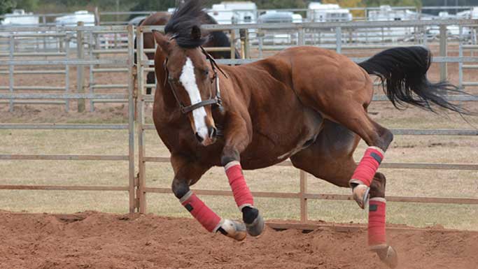
Dr. Melissa MacKinnon, a board-certified veterinary surgeon at the Milton Equine Hospital in Campbellville, ON, says that the majority of fractures referred to the hospital occur in race horses, either during a race or during high-speed training. However, sport horses and even pleasure horses do occasionally break bones while out in the pasture or during other activities.
Classifying the break
Fractures in horses are classified and can be described in combinations of the following just as they are in humans:
- Simple fractures where the bone has a single break and the bone is now in two parts;
- Comminuted fractures where the bone breaks in multiple places;
- Incomplete fractures where there are cracks in the bone, but the bone is still in one piece – more common in foals, whose bones are softer and more flexible;
- Complete fractures where the crack has completely separated two parts of the bone;
- Non-displaced fractures where the pieces of bone stay in the same position as they would have without the break;
- Displaced fractures where the bones have separated and moved apart;
- Open fractures where part of the bone breaks through the skin, which makes infection much more likely and increases the risk of complications;
- Closed fractures where the bone is not protruding through the skin;
- Stress fractures are small cracks that occur because of activity. These can become much more serious fractures if the horse is not given time to rest and heal.
MacKinnon explains that one of the most common fractures she sees is the condylar fracture – a crack or break in the horse’s cannon bone starting at the fetlock (ankle) joint and coursing up the bone. “This is definitely a race horse injury,” she says. The break can be either medial (on the inside) or lateral (on the outside), which is classified based on where the fracture line starts. MacKinnon says medial fractures often spiral up the horse’s leg, although this doesn’t always happen.
One of the most challenging aspects of these injuries is that they usually happen when the horse is moving at high speed. The horse doesn’t stop instantly when the break occurs, and each extra step he takes can cause more damage. An incomplete fracture may become complete, for example; a non-displaced fracture may become displaced.
“The incomplete and non-displaced fractures have good outcomes,” says MacKinnon. “Lateral condylar fractures can often be fixed with screws, while the medial ones sometimes require both screws and plates to fix the break.” Any condylar fracture, but especially displaced fractures, may heal, but lead to arthritis in the fetlock joint. The spiraling medial condylar fractures are unpredictable and even with appropriate surgical repair may lead to a worse fracture during recovery from general anesthesia.
Length of recovery depends on the type of condylar fracture, MacKinnon says, but a typical recommendation would be three months of stall rest, followed by a month or two of light work, and then a return to work four or five months after the injury.
Another common fracture MacKinnon sees is a break in the first phalanx (P1) of the pastern, one of two bones in the pastern (the second phalanx is referred to as P2). MacKinnon says she sees P1 fractures more often in Standardbreds racing in harness than Thoroughbred race horses, but she does encounter them in the latter, too. “These usually happen during training and racing,” she adds, “but I do sometimes see them in horses who have been turned out in pasture and injure themselves there.”
The fracture can be a short line in the centre of the bone, a spiral fracture, or in some cases involving several different planes. “The fracture can be so painful that the horse can’t put any weight on the leg at all,” she says.
Repair and recovery
Many of these fractures can be repaired with screws to hold the bone together, but some of the more complicated and comminuted ones require a transfixation pin cast. This type of cast will support some of the weight of the animal while the fracture is healing. Most can return to training in four to six months but, as with other fractures, there is a risk of arthritis developing at the site of the injury in the fetlock or pastern that might limit the horse’s ability to race or compete again. Horses with complicated fractures are generally saved for breeding or retirement.
Fractures of the second phalanx (P2) are more often seen in Western performance horses and in the hind legs because of the sliding stops and turning they are required to do. These can be treated with screws and sometimes plates.
MacKinnon also sees some fractures of the third phalanx (P3) which is actually inside the horse’s hoof. Often these are treated with a bar shoe, which restricts expansion of the hoof wall. “The hoof and the shoe act like a cast, immobilizing the fracture enough to heal,” she explains.
The horse will generally require six to eight months of stall rest with the bar shoe on. Ocassionally, the break heals with fibrous tissue rather than bone, so the crack is still visible on x-ray, but the horse is sound. In some cases a screw may be placed in P3 through the hoof wall to stabilize the fracture.
MacKinnon says that for many years – and still today – veterinarians have relied on radiographs (x-rays) taken at several different angles to help them visualize the injury and plan how to repair the fracture. Now CT scans (computed tomography) can provide a 3D image of the fracture and make it much easier to fully understand. While they are not available at all equine hospitals, CT scanners are becoming more common. Some veterinary hospitals even have small CT units that can be used in the surgery suite while the veterinarian is working on the horse, helping to guide the placement of screws and other aspects of the repair.
Another improvement which has now become commonly used is the locking compression plate with screws that thread into the plate, making the entire system stronger. “With these, veterinarians are able to use shorter plates,” MacKinnon says. The plates are often placed in a less-invasive fashion through stab incisions.
When accidents happen at home
A common pasture injury is a break to the splint bone, often caused by a kick from another horse. There are two splint bones in each leg, lying on either side of the cannon bone. In older horses these often become fused to the cannon bone, but they are more separate in younger horses. MacKinnon says that when these fracture in the bottom portion of the bone, the simplest route to healing is often to remove the fractured portion of the bone, as they often heal slowly or poorly. “If you leave it, the horse often develops a large callus around the break,” she adds.
Kicks from pasturemates can also cause breaks to the elbow part of the ulnar bone (the olecranon). When this happens, the horse will often be bent over and unable to fix his knee so that he can stand normally, because the triceps muscle is attached to this bone. The leg may be dragging as well.
“A complication with this type of fracture is that often the force of the kick has also broken the skin, so infection of the repair is a risk,” says MacKinnon. Often this is treated with a bone plate over the area to keep the bones in place as the fracture heals. In younger horses, the plate will often need to be removed after the fracture has healed in order to allow normal growth, but in older horses the plate can often be left in. In this surgery, as in all fractures repaired surgically, antibiotics will also be given to prevent infection from setting in.
Horses can also injure themselves if they bang their hips against a door frame or other solid opening, leading to a pelvic fracture of the tuber coxae of the ilium (knockdown hip). “Be careful when leading your horse in and out of the barn or stall,” says MacKinnon, noting that sometimes this happens with horses at pasture when going in and out of run-in shelters as well. This is commonly treated conservatively with sufficient rest, although it often results in a cosmetic blemish.
A special job for specialists
MacKinnon feels that it is important for owners and trainers to know that the American College of Veterinary Surgeons oversees the training and certifying of board-certified specialists in surgery, either large animal or small animal. “This work is not simple, and you really don’t learn enough during veterinary college,” she says. “To become a veterinary surgeon, you need to first graduate vet school, then do an internship, then a three-year residency and meet all of the requirements before you are able to write the specialty surgery board examination. This gives you the expertise to deal with the many different types of fractures we see in horses.”